Availability
- Request Lead Time
- In stock and ready for quick dispatch
- Usually dispatched within 5-10 working days
Product Overview
| Product Name | Aspartoacylase antibody [N1C3-2] |
|---|---|
| Catalog Number | GRP138 |
| Species/Host | Rabbit |
| Reactivity | Human, Mouse, Monkey |
| Conjugation | Unconjugated |
| Tested applications | ICC, IF, IHC-P, WB |
| Immunogen | Recombinant protein encompassing a sequence within the center region of human Aspartoacylase. The exact sequence is proprietary. |
| Alternative Names | (click to expand) |
Product Properties
| Form/Appearance | Liquid: 1XPBS, 20% Glycerol (pH7). 0.025% ProClin 300 was added as a preservative. |
|---|---|
| Concentration | 1.22 mg/ml |
| Storage | Store as concentrated solution. Centrifuge briefly prior to opening vial. For short-term storage (1-2 weeks), store at 4°C. For long-term storage, aliquot and store at -20°C or below. Avoid multiple freeze-thaw cycles. |
| Note | For research use only. |
| Isotype | IgG |
| Clonality | Polyclonal |
| Purity | Purified by antigen-affinity chromatography. |
| Uniprot ID | P45381 |
| Entrez | 443 |
Product Description
This gene encodes an enzyme that catalyzes the conversion of N-acetyl_L-aspartic acid (NAA) to aspartate and acetate. NAA is abundant in the brain where hydrolysis by aspartoacylase is thought to help maintain white matter. This protein is an NAA scavenger in other tissues. Mutations in this gene cause Canavan disease. Alternatively spliced transcript variants have been found for this gene. [provided by RefSeq]
Application Notes
| Dilution Range | WB: 1:5000-1:20000,ICC: 1:100-1:1000,IHC-P: 1:100-1:1000 |
|---|
Validation Images
![Non-transfected (–) and transfected (+) 293T whole cell extracts (30 μg) were separated by 10% SDS-PAGE, and the membrane was blotted with Aspartoacylase antibody [N1C3-2] (GRP590) diluted at 1:10000. The HRP-conjugated anti-rabbit IgG antibody was](https://www.grp-ak.de/media/catalog/product/a/s/aspartoacylase-antibody-n1c3-2_grp590_wb_4_2.jpg)
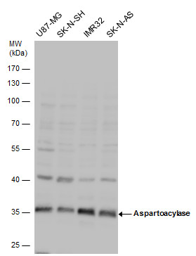
![Various whole cell extracts (30 μg) were separated by 10% SDS-PAGE, and the membrane was blotted with Aspartoacylase antibody [N1C3-2] (GRP590) diluted at 1:1000. The HRP-conjugated anti-rabbit IgG antibody was used to detect the primary antibody.](https://www.grp-ak.de/media/catalog/product/a/s/aspartoacylase-antibody-n1c3-2_grp590_wb_2_2.jpg)
![Various tissue extracts (50 μg) were separated by 10% SDS-PAGE, and the membrane was blotted with Aspartoacylase antibody [N1C3-2] (GRP590) diluted at 1:1000. The HRP-conjugated anti-rabbit IgG antibody was used to detect the primary antibody.](https://www.grp-ak.de/media/catalog/product/a/s/aspartoacylase-antibody-n1c3-2_grp590_wb_1_2.jpg)
![Aspartoacylase antibody [N1C3-2] detects Aspartoacylase protein at cytoplasm by immunofluorescent analysis.Sample: HeLa cells were fixed in 4% paraformaldehyde at RT for 15 min.Green: Aspartoacylase protein stained by Aspartoacylase antibody [N1C3-2] (GRP](https://www.grp-ak.de/media/catalog/product/a/s/aspartoacylase-antibody-n1c3-2_grp590_if_1_2.jpg)
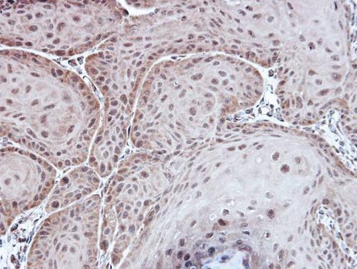
Immunohistochemical analysis of paraffin-embedded Cal27 xenograft, using Aspartoacylase(GRP590) antibody at 1:100 dilution.
Non-transfected (–) and transfected (+) 293T whole cell extracts (30 μg) were separated by 10% SDS-PAGE, and the membrane was blotted with Aspartoacylase antibody [N1C3-2] (GRP590) diluted at 1:10000. The HRP-conjugated anti-rabbit IgG antibody was
Aspartoacylase antibody detects Aspartoacylase protein by Western blot analysis. Various whole cell extracts (30 ?g) were separated by 10% SDS-PAGE, and the membrane was blotted with Aspartoacylase antibody (GRP590) diluted by 1:1000.
Various whole cell extracts (30 μg) were separated by 10% SDS-PAGE, and the membrane was blotted with Aspartoacylase antibody [N1C3-2] (GRP590) diluted at 1:1000. The HRP-conjugated anti-rabbit IgG antibody was used to detect the primary antibody.
Various tissue extracts (50 μg) were separated by 10% SDS-PAGE, and the membrane was blotted with Aspartoacylase antibody [N1C3-2] (GRP590) diluted at 1:1000. The HRP-conjugated anti-rabbit IgG antibody was used to detect the primary antibody.
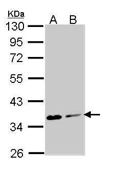
Sample (30 μg of whole cell lysate) A: Molt-4 (GRP590) B: Raji 10% SDS PAGE diluted at 1:1000 The HRP-conjugated anti-rabbit IgG antibody was used to detect the primary antibody.
Aspartoacylase antibody [N1C3-2] detects Aspartoacylase protein at cytoplasm by immunofluorescent analysis.Sample: HeLa cells were fixed in 4% paraformaldehyde at RT for 15 min.Green: Aspartoacylase protein stained by Aspartoacylase antibody [N1C3-2] (GRP
Reviews
Write Your Own Review
