Availability
- Request Lead Time
- In stock and ready for quick dispatch
- Usually dispatched within 5-10 working days
Product Overview
| Product Name | E-Cadherin antibody |
|---|---|
| Catalog Number | GRP7 |
| Species/Host | Rabbit |
| Reactivity | Human, Mouse, Rat, Zebrafish |
| Conjugation | Unconjugated |
| Tested applications | ICC, IF, IHC-P, IP, WB |
| Immunogen | Recombinant protein encompassing a sequence within the center region of human E-Cadherin. The exact sequence is proprietary. |
| Alternative Names | (click to expand) |
Product Properties
| Form/Appearance | Liquid: 1XPBS, 1% BSA, 20% Glycerol (pH7). 0.025% ProClin 300 was added as a preservative. |
|---|---|
| Concentration | 0.49 mg/ml |
| Storage | Store as concentrated solution. Centrifuge briefly prior to opening vial. For short-term storage (1-2 weeks), store at 4°C. For long-term storage, aliquot and store at -20°C or below. Avoid multiple freeze-thaw cycles. |
| Note | For research use only. |
| Isotype | IgG |
| Clonality | Polyclonal |
| Purity | Purified by antigen-affinity chromatography. |
| Uniprot ID | P12830 |
| Entrez | 999 |
Product Description
This gene is a classical cadherin from the cadherin superfamily. The encoded protein is a calcium dependent cell-cell adhesion glycoprotein comprised of five extracellular cadherin repeats, a transmembrane region and a highly conserved cytoplasmic tail. Mutations in this gene are correlated with gastric, breast, colorectal, thyroid and ovarian cancer. Loss of function is thought to contribute to progression in cancer by increasing proliferation, invasion, and/or metastasis. The ectodomain of this protein mediates bacterial adhesion to mammalian cells and the cytoplasmic domain is required for internalization. Identified transcript variants arise from mutation at consensus splice sites. [provided by RefSeq]
Application Notes
| Dilution Range | WB: 1:500-1:10000,ICC: 1:100-1:1000,IHC-P: 1:100-1:1000,IP: 1:100-1:500 |
|---|
Validation Images
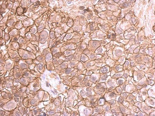
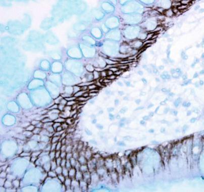
E-cadherin antibody detects E-cadherin protein at membrane on human breast cancer by immunohistochemical analysis. Sample: Paraffin-embedded breast cancer. E-cadherin antibody (GRP459) dilution: 1:500.
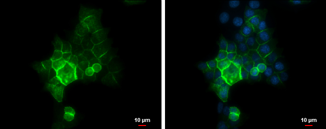
E-Cadherin antibody detects E-Cadherin protein at cell membrane by immunofluorescent analysis.Sample: HCT 116 cells were fixed in 4% paraformaldehyde at RT for 15 min.Green: E-Cadherin protein stained by E-Cadherin antibody (GRP459) diluted at 1:500.Blue:
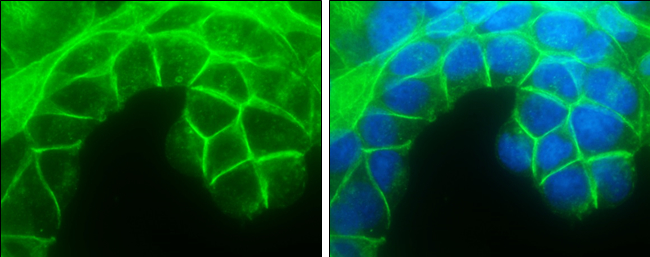
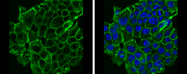
E-Cadherin antibody detects E-Cadherin protein at cell membrane by immunofluorescent analysis.Sample: MCF7 cells were fixed in 4% paraformaldehyde at RT for 15 min.Green: E-Cadherin protein stained by E-Cadherin antibody (GRP459) diluted at 1:500.Blue: Ho
E-Cadherin antibody detects E-Cadherin protein at cell membrane in mouse cervix by immunohistochemical analysis. Sample: Paraffin-embedded mouse cervix. E-Cadherin antibody (GRP459) diluted at 1:500.
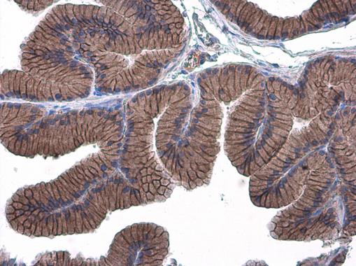
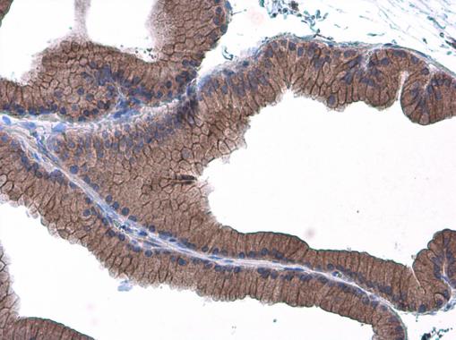
E-Cadherin antibody detects E-Cadherin protein at cell membrane in rat prostate by immunohistochemical analysis. Sample: Paraffin-embedded rat prostate. E-Cadherin antibody (GRP459) diluted at 1:500.
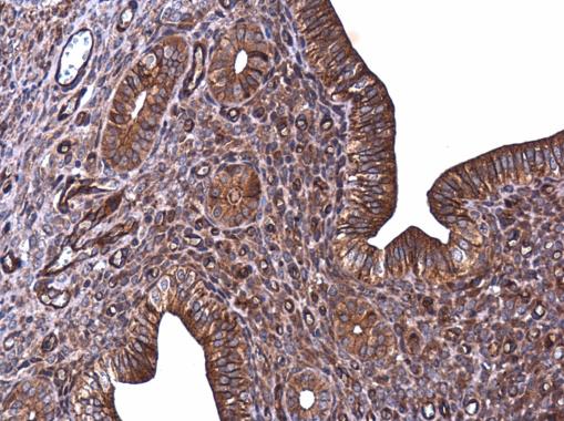
Immunohistochemical analysis of paraffin-embedded human ulcerative colitis tissue using E-Cadherin antibody (GRP459).
E-Cadherin antibody detects E-Cadherin protein at cell membrane in mouse pancreas by immunohistochemical analysis. Sample: Paraffin-embedded mouse pancreas. E-Cadherin antibody (GRP459) diluted at 1:400.
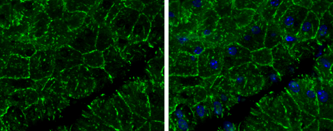
E-Cadherin antibody detects E-Cadherin protein at cell membrane by immunofluorescent analysis.Sample: A431 cells were fixed in 4% paraformaldehyde at RT for 15 min.Green: E-Cadherin protein stained by E-Cadherin antibody (GRP459) diluted at 1:500.Blue: Ho
E-Cadherin antibody detects E-Cadherin protein at cell membrane in rat prostate by immunohistochemical analysis. Sample: Paraffin-embedded rat prostate. E-Cadherin antibody (GRP459) diluted at 1:500.
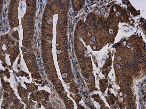
E-Cadherin antibody detects E-Cadherin protein at cell membrane and cytoplasm in rat duodenum by immunohistochemical analysis. Sample: Paraffin-embedded rat duodenum. E-Cadherin antibody (GRP459) diluted at 1:500.
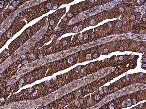
E-Cadherin antibody detects E-Cadherin protein at cell membrane in rat intestine by immunohistochemical analysis. Sample: Paraffin-embedded rat intestine. E-Cadherin antibody (GRP459) diluted at 1:500.
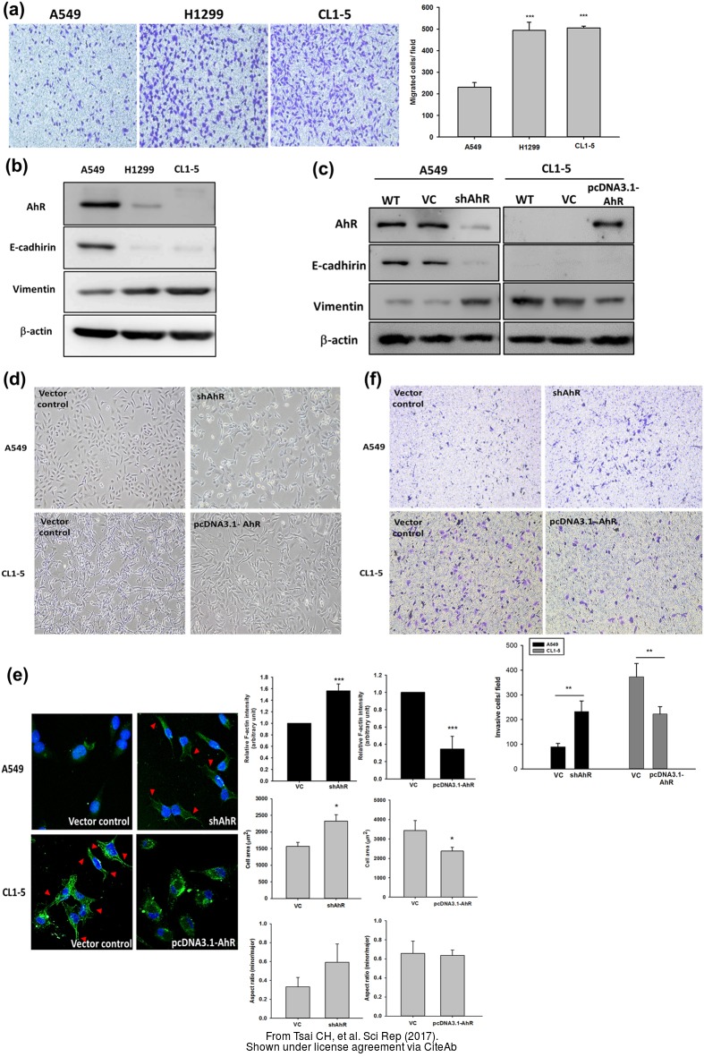
The WB analysis of E-Cadherin antibody was published by Tsai CH and colleagues in the journal Sci Rep in 2017.PMID: 28195146
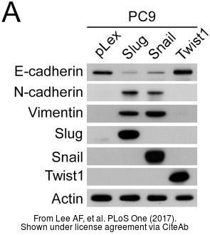
The WB analysis of E-Cadherin antibody was published by Lee AF and colleagues in the journal PLoS One in 2017.PMID: 28683123
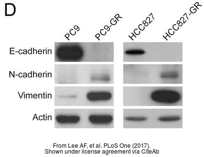
The WB analysis of E-Cadherin antibody was published by Lee AF and colleagues in the journal PLoS One in 2017.PMID: 28683123
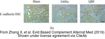
The IHC-P analysis of E-Cadherin antibody was published by Zhang X and colleagues in the journal Evid Based Complement Alternat Med in 2019.PMID: 31186661
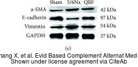
The WB analysis of E-Cadherin antibody was published by Zhang X and colleagues in the journal Evid Based Complement Alternat Med in 2019.PMID: 31186661
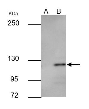
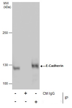
E-cadherin antibody immunoprecipitates E-cadherin protein in IP experiments.IP samples: MCF-7 whole cell extractA. Control with 3 ?g of preimmune Rabbit IgGB. Immunoprecipitation of E-cadherin protein by 3 ?g E-cadherin antibody (GRP459)5 % SDS-PAGEThe im
Reviews
Write Your Own Review

