Availability
- Request Lead Time
- In stock and ready for quick dispatch
- Usually dispatched within 5-10 working days
Histone H2A.XS139ph (phospho Ser139) antibody
Catalog Number: GRP67
Product Overview
| Product Name | Histone H2A.XS139ph (phospho Ser139) antibody |
|---|---|
| Catalog Number | GRP67 |
| Species/Host | Rabbit |
| Reactivity | Human, Mouse, Rat, Zebrafish |
| Conjugation | Unconjugated |
| Tested applications | ICC, IF, IHC-P, WB |
| Immunogen | Carrier-protein conjugated synthetic peptide corresponding to residues around human Histone H2A.XS139ph (phospho Ser139). The exact sequence is proprietary. |
| Alternative Names | (click to expand) |
Product Properties
| Form/Appearance | Liquid: 1XPBS, 1% BSA, 20% Glycerol (pH7). 0.025% ProClin 300 was added as a preservative. |
|---|---|
| Concentration | 0.16 mg/ml |
| Storage | Store as concentrated solution. Centrifuge briefly prior to opening vial. For short-term storage (1-2 weeks), store at 4°C. For long-term storage, aliquot and store at -20°C or below. Avoid multiple freeze-thaw cycles. |
| Note | For research use only. |
| Isotype | IgG |
| Clonality | Polyclonal |
| Purity | Purified by antigen-affinity chromatography. |
| Uniprot ID | P16104 |
| Entrez | 3014 |
Product Description
Histones are basic nuclear proteins that are responsible for the nucleosome structure of the chromosomal fiber in eukaryotes. Two molecules of each of the four core histones (H2A, H2B, H3, and H4) form an octamer, around which approximately 146 bp of DNA is wrapped in repeating units, called nucleosomes. The linker histone, H1, interacts with linker DNA between nucleosomes and functions in the compaction of chromatin into higher order structures. This gene encodes a member of the histone H2A family, and generates two transcripts through the use of the conserved stem-loop termination motif, and the polyA addition motif. [provided by RefSeq]
Application Notes
| Dilution Range | WB: 1:500-1:3000,ICC: 1:100-1:1000,IHC-P: 1:100-1:1000 |
|---|
Validation Images
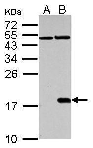
Histone H2A.X (phospho Ser139) antibody detects H2AFX protein in cisplatin-treated samples by western blot analysis.A. 30 μg NIH-3T3 whole cell lysate/extract (untreated) B. 30 μg NIH-3T3 whole cell lysate/extract (30μM cisplatin treatment for 24
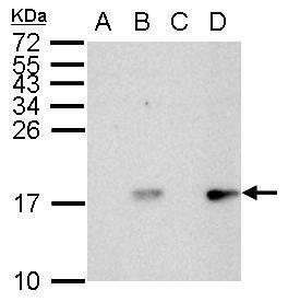
Histone H2A.X (phospho Ser139) antibody detects H2AFX protein by western blot analysis.A. 30 μg HCT116 whole cell lysate/extract (untreated for 2hr)B. 30 μg HCT116 whole cell lysate/extract (UVB treatment 50mJ for 2hr)C. 30 μg HCT116 whole cell l
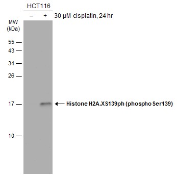
Untreated (–) and treated (+) HCT-116 whole cell extract (30 μg) were separated by 15% SDS-PAGE, and the membrane was blotted with Histone H2A.XS139ph (phospho Ser139) antibody (GRP519) diluted at 1:1000. The HRP-conjugated anti-rabbit IgG antibody
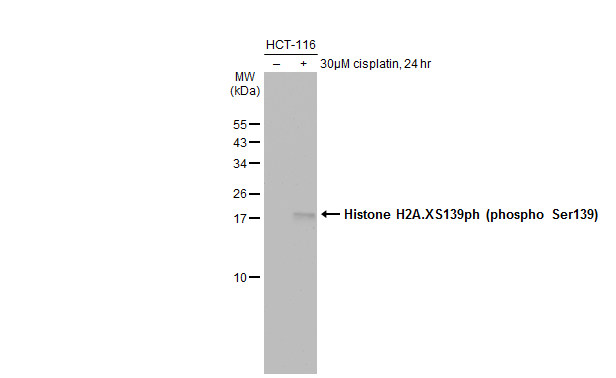
Untreated (–) and treated (+) HCT-116 whole cell extracts (30 μg) were separated by 15% SDS-PAGE, and the membrane was blotted with Histone H2A.XS139ph (phospho Ser139) antibody (GRP519) diluted at 1:1000. The HRP-conjugated anti-rabbit IgG antibody
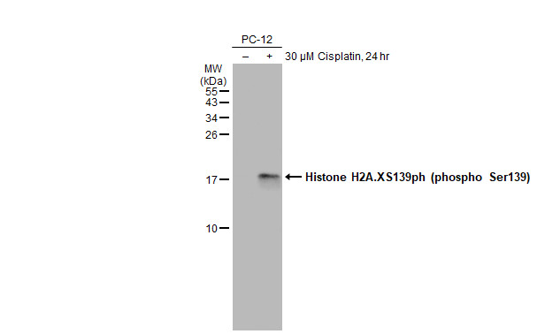
Untreated (–) and treated (+) PC-12 whole cell extracts (30 μg) were separated by 15% SDS-PAGE, and the membrane was blotted with Histone H2A.XS139ph (phospho Ser139) antibody (GRP519) diluted at 1:1000. The HRP-conjugated anti-rabbit IgG antibody w
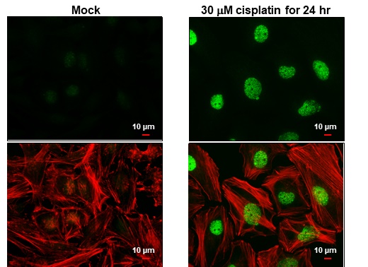
Histone H2A.XS139ph (phospho Ser139) antibody detects Histone H2A.XS139ph (phospho Ser139) protein at nucleus by immunofluorescent analysis.Sample: HeLa cells were fixed in 4% paraformaldehyde at RT for 15 min.Green: Histone H2A.XS139ph (phospho Ser139) s
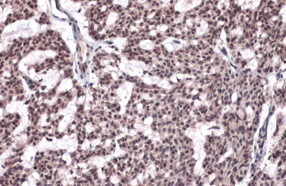
Histone H2A.XS139ph (phospho Ser139) antibody detects Histone H2A.XS139ph (phospho Ser139) protein at nucleus by immunohistochemical analysis.Sample: Paraffin-embedded human breast carcinoma.Histone H2A.XS139ph (phospho Ser139) stained by Histone H2A.XS13
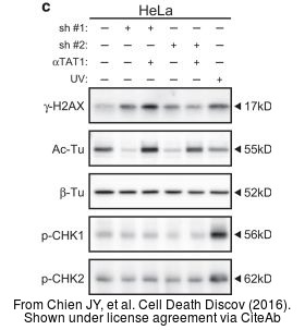
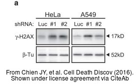
The WB analysis of Histone H2A.XS139ph (phospho Ser139) antibody was published by Chien JY and colleagues in the journal Cell Death Discov in 2016.PMID: 27551500
Reviews
Write Your Own Review
