Availability
- Request Lead Time
- In stock and ready for quick dispatch
- Usually dispatched within 5-10 working days
Product Overview
| Product Name | LDHA antibody |
|---|---|
| Catalog Number | GRP21 |
| Species/Host | Rabbit |
| Reactivity | Human, Mouse, Rat |
| Conjugation | Unconjugated |
| Tested applications | ICC, IF, IHC-P, WB |
| Immunogen | Recombinant protein encompassing a sequence within the center region of human LDHA. The exact sequence is proprietary. |
| Alternative Names | (click to expand) |
Product Properties
| Form/Appearance | Liquid: 1XPBS, 1% BSA, 20% Glycerol (pH7). 0.025% ProClin 300 was added as a preservative. |
|---|---|
| Concentration | 0.48 mg/ml |
| Storage | Store as concentrated solution. Centrifuge briefly prior to opening vial. For short-term storage (1-2 weeks), store at 4°C. For long-term storage, aliquot and store at -20°C or below. Avoid multiple freeze-thaw cycles. |
| Note | For research use only. |
| Isotype | IgG |
| Clonality | Polyclonal |
| Purity | Purified by antigen-affinity chromatography. |
| Uniprot ID | P00338 |
| Entrez | 3939 |
Product Description
The protein encoded by this gene catalyzes the conversion of L-lactate and NAD to pyruvate and NADH in the final step of anaerobic glycolysis. The protein is found predominantly in muscle tissue and belongs to the lactate dehydrogenase family. Mutations in this gene have been linked to exertional myoglobinuria. Multiple transcript variants encoding different isoforms have been found for this gene. The human genome contains several non-transcribed pseudogenes of this gene. [provided by RefSeq]
Application Notes
| Dilution Range | WB: 1:500-1:3000,ICC: 1:100-1:1000,IHC-P: 1:100-1:1000 |
|---|
Validation Images
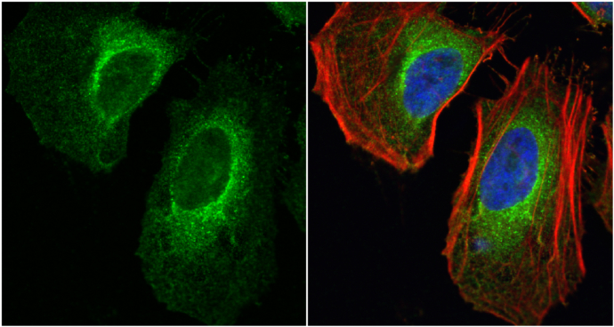
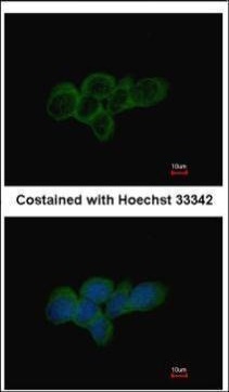
LDHA antibody detects LDHA protein at cytoplasm by immunofluorescent analysis.Sample: HeLa cells were fixed in 4% paraformaldehyde at RT for 15 min.Green: LDHA protein stained by LDHA antibody (GRP473) diluted at ) diluted at 1:200.Blue: Hoechst 33342 sta
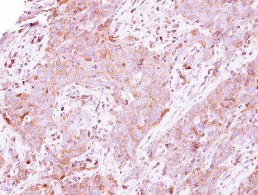
Immunohistochemical analysis of paraffin-embedded human lung papillory adenocarcinoma, using LDHA(GRP473) antibody at 1:500 dilution.
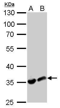
LDHA antibody detects LDHA protein by western blot analysis.A. 30 μg PC-12 whole cell lysate/extractB. 30 μg Rat2 whole cell lysate/extract10% SDS-PAGELDHA antibody (GRP473) dilution: 1:1000 The HRP-conjugated anti-rabbit IgG antibody was used to d
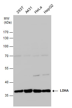
LDHA antibody detects LDHA protein by western blot analysis. Various whole cell extracts (30 μg) were separated by 10% SDS-PAGE, and the membrane was blotted with LDHA antibody (GRP473) diluted at 1:1000. The HRP-conjugated anti-rabbit IgG antibody wa
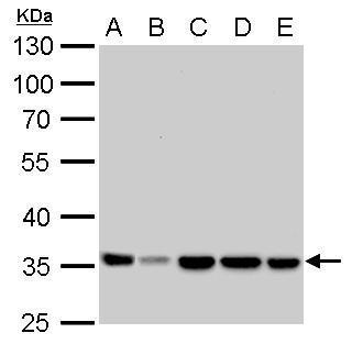
LDHA antibody detects LDHA protein by western blot analysis.A. 30 μg Neuro2A whole cell lysate/extract B. 30 μg C8D30 whole cell lysate/extract C. 30 μg NIH-3T3 whole cell lysate/extract D. 30 μg Raw264.7 whole cell lysate/extract E. 30 μg
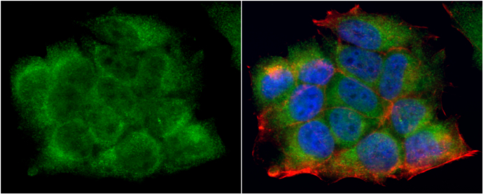
LDHA antibody detects LDHA protein at cytoplasm by immunofluorescent analysis.Sample: MCF7 cells were fixed in 4% paraformaldehyde at RT for 15 min.Green: LDHA protein stained by LDHA antibody (GRP473) diluted at ) diluted at 1:200.Blue: Hoechst 33342 sta
Immunofluorescence analysis of methanol-fixed A431, using LDHA(GRP473) antibody at 1:500 dilution.
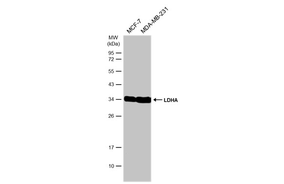
Various whole cell extracts (30 μg) were separated by 12% SDS-PAGE, and the membrane was blotted with LDHA antibody (GRP473) diluted at 1:1000. The HRP-conjugated anti-rabbit IgG antibody was used to detect the primary antibody.
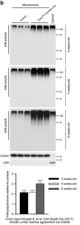
The WB analysis of LDHA antibody was published by Ugun-Klusek A and colleagues in the journal Cell Death Dis in 2017.PMID: 28055010
Reviews
Write Your Own Review
