Availability
- Request Lead Time
- In stock and ready for quick dispatch
- Usually dispatched within 5-10 working days
Product Overview
| Product Name | TDP43 antibody |
|---|---|
| Catalog Number | GRP141 |
| Species/Host | Rabbit |
| Reactivity | Human, Mouse, Rat |
| Conjugation | Unconjugated |
| Tested applications | ChIP, ICC, IF, IHC-P, IP, WB |
| Immunogen | Recombinant protein encompassing a sequence within the center region of human TDP43. The exact sequence is proprietary. |
| Alternative Names | (click to expand) |
Product Properties
| Form/Appearance | Liquid: 1XPBS, 20% Glycerol (pH7). 0.025% ProClin 300 was added as a preservative. |
|---|---|
| Concentration | 1.1 mg/ml |
| Storage | Store as concentrated solution. Centrifuge briefly prior to opening vial. For short-term storage (1-2 weeks), store at 4°C. For long-term storage, aliquot and store at -20°C or below. Avoid multiple freeze-thaw cycles. |
| Note | For research use only. |
| Isotype | IgG |
| Clonality | Polyclonal |
| Purity | Purified by antigen-affinity chromatography. |
| Uniprot ID | Q13148 |
| Entrez | 23435 |
Product Description
HIV-1, the causative agent of acquired immunodeficiency syndrome (AIDS), contains an RNA genome that produces a chromosomally integrated DNA during the replicative cycle. Activation of HIV-1 gene expression by the transactivator Tat is dependent on an RNA regulatory element (TAR) located downstream of the transcription initiation site. The protein encoded by this gene is a transcriptional repressor that binds to chromosomally integrated TAR DNA and represses HIV-1 transcription. In addition, this protein regulates alternate splicing of the CFTR gene. A similar pseudogene is present on chromosome 20. [provided by RefSeq]
Application Notes
| Dilution Range | WB: 1:500-1:3000,ICC: 1:100-1:1000,IHC-P: 1:100-1:1000,IP: 1:100-1:1000 |
|---|
Validation Images
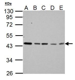
TARDBP antibody detects TARDBP protein by western blot analysis.A. 30 μg Neuro2A whole cell lysate/extractB. 30 μg GL261 whole cell lysate/extractC. 30 μg NIH-3T3 whole cell lysate/extractD. 30 μg BCL-1 whole cell lysate/extractE. 30 μg Raw
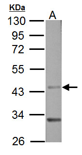
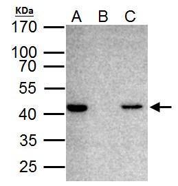
Sample (50 μg of whole cell lysate) A: Rat brain 10% SDS PAGE GRP593 diluted at 1:3000 The HRP-conjugated anti-rabbit IgG antibody was used to detect the primary antibody.
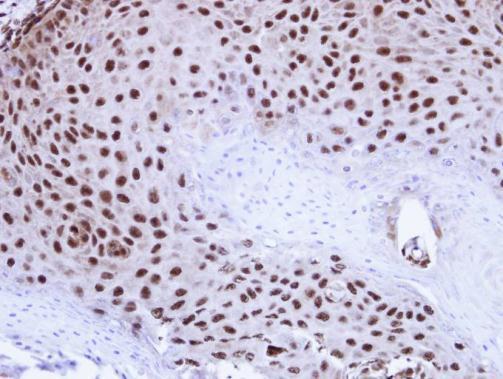
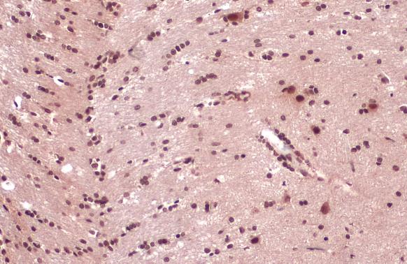
Immunohistochemical analysis of paraffin-embedded Cal27 Xenograft , using TDP-43(GRP593) antibody at 1:100 dilution.
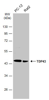
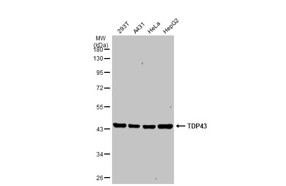
Various whole cell extracts (30 μg) were separated by 10% SDS-PAGE, and the membrane was blotted with TDP43 antibody (GRP593) diluted at 1:2000. The HRP-conjugated anti-rabbit IgG antibody was used to detect the primary antibody.
Various whole cell extracts (30 μg) were separated by 10% SDS-PAGE, and the membrane was blotted with TDP43 antibody (GRP593) diluted at 1:1000. The HRP-conjugated anti-rabbit IgG antibody was used to detect the primary antibody.
TDP43 antibody detects TDP43 protein at cytoplasm and nucleus by immunohistochemical analysis.Sample: Paraffin-embedded rat brain.TDP43 stained by TDP43 antibody (GRP593) diluted at 1:2000.Antigen Retrieval: Citrate buffer, pH 6.0, 15 min
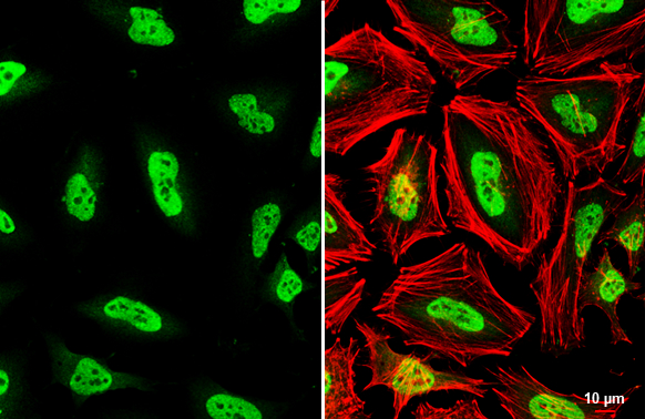
TDP43 antibody detects TDP43 protein at nucleus by immunofluorescent analysis.Sample: HeLa cells were fixed in 4% paraformaldehyde at RT for 15 min.Green: TDP43 stained by TDP43 antibody (GRP593) diluted at 1:500.Red: phalloidin, a cytoskeleton marker, di
TARDBP antibody immunoprecipitates TARDBP protein in IP experiments. IP Sample: HeLa whole cell lysate/extract A. 40 ?g HeLa whole cell lysate/extract B. Control with 2 ?g of preimmune rabbit IgG C. Immunoprecipitation of TARDBP protein by 2 ?g of TARDBP
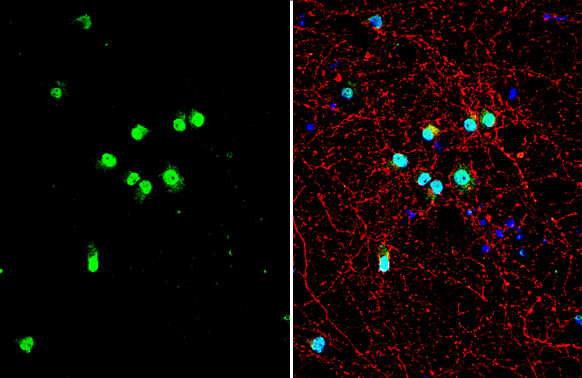
TDP43 antibody detects TDP43 protein by immunofluorescent analysis.Sample: DIV10 rat E18 primary cortical neuron cells were fixed in 4% paraformaldehyde at RT for 15 min.Green: TDP43 stained by TDP43 antibody (GRP593) diluted at 1:500.Red: Tau, stained by
Reviews
Write Your Own Review
